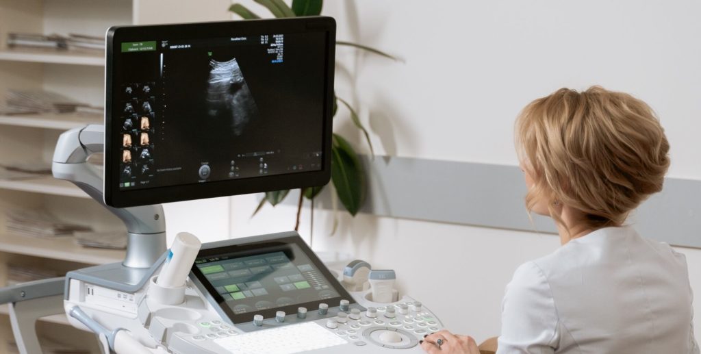
July 22, 2024

Ultrasound Machine

Ultrasound Machine

Introduction To Ultrasound Machine An ultrasound machine uses high-frequency sound waves to produce images of the inside of the body. It is commonly used for imaging soft tissues, organs, and monitoring pregnancies. Key Features: Transducer Probe: The handheld device that emits sound waves and receives the echoes that bounce back. Sound Waves: The transducer sends sound waves into the body. These waves reflect off tissues, organs, and fluids, and return to the transducer. Image Formation: The machine processes these echoes to create real-time images on a monitor. Applications: Obstetrics: For monitoring fetal development during pregnancy. Abdominal Imaging: To examine organs such as the liver, gallbladder, spleen, pancreas, and kidneys. Cardiology: Echocardiograms are specialized ultrasound tests used to assess the heart’s function and structure.

Ultrasound Machine Read More »

ECG Machine (Electrocardiogram Machine)

ECG Machine (Electrocardiogram Machine)

Introduction To ECG Machine (Electrocardiogram Machine) An ECG machine is used to record the electrical activity of the heart over a period of time. It is a non-invasive procedure that helps in diagnosing various heart conditions. Key Features: Electrodes: Small sticky patches (electrodes) are placed on the patient’s chest, arms, and legs. These electrodes are connected to the ECG machine. Electrical Activity Recording: The machine records the electrical signals that trigger heartbeats and displays them as waves on a screen or paper. Waveform Analysis: The recorded waveforms help in diagnosing arrhythmias, heart attacks, and other cardiac problems. Applications: Diagnosis of Arrhythmias: Identifies irregular heartbeats. Myocardial Infarction: Detects heart attacks by identifying abnormal wave patterns. Monitoring: Used for continuous monitoring in critical care settings.

ECG Machine (Electrocardiogram Machine) Read More »

MRI Scanner

MRI Scanner (Magnetic Resonance Imaging Scanner)

Introduction to MRI Scanner An MRI scanner is a medical imaging device used to create detailed images of the organs and tissues within the body. It uses powerful magnets, radio waves, and a computer to generate these images without using ionizing radiation (like X-rays or CT scans). Key Features: Magnetic Field: The MRI scanner uses a strong magnetic field to align the protons in the body’s water molecules. Radio Waves: Radiofrequency pulses are then used to disturb this alignment. When the protons realign with the magnetic field, they emit signals that are captured by the scanner. Computer Processing: These signals are processed by a computer to produce detailed cross-sectional images of the body. Applications: Neurology: For imaging the brain and spinal cord to diagnose conditions like tumors, stroke, and multiple sclerosis. Orthopedics: For visualizing joint and soft tissue injuries. Cardiology: For assessing heart and vascular diseases.

MRI Scanner (Magnetic Resonance Imaging Scanner) Read More »

Scroll to Top